

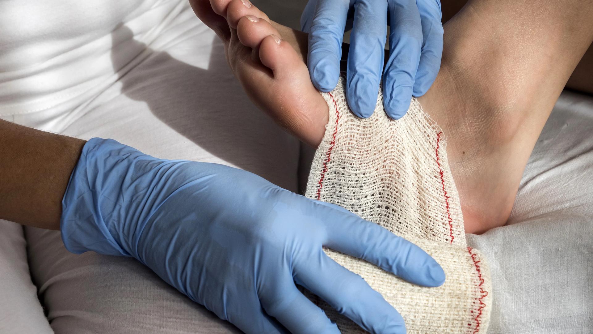
Diabetic foot ulcers develop in approximately 25 percent of people with diabetes during their lifetime, and 14 to 24 percent of those ulcers ultimately lead to lower-extremity amputation. Early, accurate assessment and aggressive multidisciplinary treatment are the single biggest levers in changing that outcome -- and yet diabetic foot ulcer management remains one of the most under-standardized areas of wound care across post-acute and outpatient settings.
DFUs are uniquely complex because they sit at the intersection of three failures: peripheral neuropathy that removes the protective pain warning, peripheral arterial disease that impairs healing physiology, and biomechanical pressure that maintains the wound. Effective management requires coordinated care across podiatry, vascular surgery, infectious disease, endocrinology, and wound care -- with documentation that follows the patient across handoffs.
This guide is a clinical reference for DFU assessment, classification, treatment, and documentation, built around current IWGDF, ADA, and IDSA guidance. It is written for wound care nurses, podiatrists, primary care clinicians, and wound program directors who manage DFUs across the continuum of care.
What Is a Diabetic Foot Ulcer?
Per IWGDF definitions, a diabetic foot ulcer is a break in the skin of the foot in a patient with diabetes mellitus, extending through the dermis at minimum, occurring below the malleoli. The DFU phenotype is distinct from other chronic wounds because three pathologies converge:
- Peripheral neuropathy. Loss of protective sensation removes the pain-driven behavioral feedback that prevents tissue damage from continued pressure. Patients walk on ulcers without realizing it.
- Peripheral arterial disease (PAD). Reduced tissue perfusion impairs every phase of healing -- inflammation, granulation, epithelialization. Without adequate flow, no other intervention restores closure.
- Infection susceptibility. Hyperglycemia, vascular compromise, and impaired neutrophil function combine to lower the threshold for both local and deep infection, including osteomyelitis.
Common ulcer locations follow the pressure distribution of the diabetic foot: plantar surface (especially metatarsal heads, heel, and great toe), interdigital spaces, and dorsum of the foot at areas of friction. Global prevalence among people with diabetes is approximately 6.3 percent at any given time, per IWGDF data.
Major risk factors include peripheral neuropathy, PAD, prior ulcer or amputation, foot deformity (notably Charcot neuroarthropathy), uncontrolled hyperglycemia, smoking, dialysis dependency, and transplant immunosuppression. The strongest single predictor of a future DFU is a prior DFU -- meaning that the post-healing surveillance window is itself a critical clinical phase.
Diabetic Foot Ulcer Classification Systems
Multiple validated classification systems exist; the two most widely used in clinical practice are Wagner and the University of Texas (UT) system. The IWGDF additionally recommends the SINBAD system for research and benchmarking.
Wagner Classification System
Wagner grades DFUs from 0 to 5 by depth and presence of gangrene. The system is older and limited in that it does not systematically address ischemia or infection, but it remains widely used in clinical documentation and billing.
- Grade 0. Pre-ulcerative or post-ulcerative lesion. Intact skin with risk factors present, often hyperkeratosis at pressure points.
- Grade 1. Superficial ulcer, partial or full thickness, no infection.
- Grade 2. Deeper ulcer extending to tendon, capsule, or bone -- *without* bone involvement, abscess, or osteomyelitis.
- Grade 3. Deep ulcer with abscess, osteomyelitis, or joint sepsis.
- Grade 4. Localized gangrene (forefoot or heel).
- Grade 5. Extensive gangrene of the entire foot.
The Wagner system's principal limitation is that two Grade 2 ulcers can have radically different prognoses depending on infection and ischemia status -- factors Wagner does not capture.
University of Texas (UT) Classification
The UT system uses a two-axis matrix: depth crossed with co-morbidity status, producing a clinically meaningful prognostic grid.
- Stages (depth): 0 (pre- or post-ulcerative), 1 (superficial, no tendon/capsule/bone involvement), 2 (penetrates to tendon or capsule), 3 (penetrates to bone or joint).
- Grades (co-morbidity): A (no infection or ischemia), B (infected), C (ischemic), D (infected *and* ischemic).
UT is generally preferred clinically because it captures the multi-factorial nature of DFU prognosis. A UT Stage 1A ulcer (superficial, clean) has dramatically different management implications and healing trajectory than a UT Stage 3D ulcer (penetrates to bone, both infected and ischemic), even though both are "DFU."
IWGDF / SINBAD Classification
SINBAD scores six domains: Site, Ischemia, Neuropathy, Bacterial infection, Area, and Depth. Each domain is scored 0 or 1, producing a total score from 0 to 6. The IWGDF recommends SINBAD for research, registry, and quality benchmarking applications because it is simple to apply, captures clinically relevant variables, and produces a single comparable number across settings.
Comprehensive DFU Assessment
A complete DFU assessment integrates history, multi-system physical exam, structured wound evaluation, and targeted imaging.
Patient history. Diabetes duration and current control (most recent HbA1c). Prior ulcer history, prior amputations, Charcot history. Smoking status. Dialysis or transplant status. Immunosuppressant medications. Footwear and weight-bearing patterns.
Physical examination. Visual inspection for deformity, callus, pressure-point patterns, and ulcer characteristics. Neurologic exam using the 10-gram Semmes-Weinstein monofilament (loss at any of the standard plantar test sites indicates loss of protective sensation), 128 Hz vibration tuning fork, and Achilles reflex. Vascular exam includes pedal pulse palpation, capillary refill, ankle-brachial index (ABI), and toe pressures or transcutaneous oxygen measurement (TcPO2) when ABI is unreliable from medial calcification. Musculoskeletal exam for foot deformity, range of motion, and gait abnormalities.
Wound assessment. Location and dimensions (length x width x depth) measured with a standardized technique. Probe-to-bone test for bone involvement. Wound bed characterization (percentage granulation, slough, eschar, exposed structures). Exudate volume and character. Odor. Periwound assessment for erythema, warmth, induration, and surrounding callus. Infection signs per PEDIS or IDSA criteria, recognizing that diabetic patients may not mount typical inflammatory responses.
Imaging when indicated. Plain X-ray as first-line for bone involvement, gas, foreign body, and osteomyelitis screening. MRI as the imaging gold standard for osteomyelitis when X-ray is equivocal. Duplex ultrasound, CT angiography, or formal angiography in patients who are revascularization candidates based on clinical or ABI findings.
Diabetic Foot Ulcer Treatment Framework
DFU management rests on three pillars. Skipping or under-executing any one of them is the most common cause of treatment failure.
Pillar 1 -- Offloading
Pressure is what created and maintains the ulcer. Without offloading, no debridement protocol, no advanced therapy, and no antibiotic regimen will produce sustained healing. Offloading is also the single most under-implemented pillar in real-world practice.
- Total contact casting (TCC). The clinical gold standard for plantar neuropathic ulcers. Reduces peak plantar pressure by approximately 84 percent and produces faster healing rates than most alternatives in head-to-head trials.
- Removable cast walkers. Effective when used as prescribed; rendering them irremovable (with a fiberglass overwrap or other technique) substantially improves outcomes by removing patient adherence as a variable.
- Therapeutic footwear and custom orthotics. Primarily for prevention and post-healing maintenance, not for active large-ulcer treatment.
The most common pitfall: prescribing offloading but not verifying use. Patient-reported adherence overestimates actual offloading time.
Pillar 2 -- Vascular Optimization
Adequate perfusion is the precondition for healing. ABI less than 0.9 suggests PAD; toe pressures less than 30 mmHg predict very poor spontaneous healing and trigger revascularization evaluation. TcPO2 less than 30 mmHg correlates with non-healing.
Vascular optimization includes timely revascularization referral for critical limb ischemia (endovascular or surgical bypass per the vascular team's call), glycemic control as an adjunct (ADA Standards of Care target HbA1c generally less than 7-8 percent, individualized), and smoking cessation. Smoking cessation often produces measurable healing improvements within months.
Pillar 3 -- Wound Bed Management and Infection Control
Sharp surgical debridement is preferred for most DFUs to remove callus, slough, biofilm, and devitalized tissue at each visit. Aggressive debridement at each visit is associated with faster healing than less-frequent debridement in retrospective analyses.
Bioburden management uses antimicrobial dressings when critical colonization or local infection is present. Systemic antibiotics are reserved for clinically infected wounds per IDSA criteria, tailored to severity (mild, moderate, or severe). Specific antibiotic regimens should follow current IDSA Diabetic Foot Infection guidelines and local antibiogram data.
Osteomyelitis typically requires both surgical debridement of infected bone and prolonged antibiotic therapy (typically 4 to 6 weeks per IDSA), with regimens individualized to organism and host factors.
For wounds that do not show at least 50 percent area reduction at 4 weeks of adequate standard care, advanced therapies should be considered: cellular and tissue-based products (CTPs), negative pressure wound therapy (NPWT) for appropriate wound types, hyperbaric oxygen therapy in select cases meeting CMS coverage criteria, and growth factors (becaplermin, PDGF). These therapies are escalation tools, not first-line substitutes for the three pillars.
When to Escalate Care
A multidisciplinary referral threshold lowers the risk of preventable amputation. Triggers include:
- Suspected or confirmed osteomyelitis.
- Critical limb ischemia (rest pain, gangrene, ABI less than 0.4).
- Non-healing wound at 4 weeks despite documented adequate offloading, debridement, and infection control.
- Charcot neuroarthropathy, acute or chronic.
- Severe infection requiring inpatient or surgical management.
The diabetic foot team model -- podiatry, vascular, infectious disease, endocrinology, wound care, and prosthetics, coordinated through a unified care platform -- has consistent evidence supporting reduced amputation rates compared with fragmented care models. The operational challenge is that these teams rarely sit in the same building; documentation and photo-based handoffs are what allow them to coordinate care across days, sites, and shifts.
Documentation and Coding for Diabetic Foot Ulcers
ICD-10 specificity matters more for DFU than for almost any other wound category, because reimbursement depends on combination coding that documents the diabetes type, foot ulcer presence, laterality, severity, and any complicating conditions.
Required ICD-10 components.
- E11.621 (Type 2 diabetes with foot ulcer) or E10.621 (Type 1 diabetes with foot ulcer). E11.622 / E10.622 cover other skin ulcers.
- L97.4xx and L97.5xx -- non-pressure chronic ulcer of foot, laterality- and severity-specific.
- M86.xxx -- osteomyelitis codes, added when documented.
Common CPT codes used in DFU care include debridement (97597 and 97598 for selective; 11042 through 11047 for surgical depth-specific), application of cellular and tissue-based products (15271 through 15278), and NPWT (97605 through 97608). The full CPT reference is covered in a separate post (see internal links).
The documentation-to-coding linkage is where claims live or die. ICD-10 codes must be supported by explicit documentation of neuropathy, ischemia, ulcer depth, infection status, and laterality. Templates that prompt clinicians for each required element at point of care prevent the most common denial cause: documentation that does not support the level of code billed.
V3 Biomedical's wound care platform provides specialty-specific DFU templates with structured prompts for neuropathy and vascular findings, integrated calibrated photography with measurement, and automatic alignment between documented findings and the ICD-10 / CPT codes those findings support. This is the documentation infrastructure that turns clinical assessment into defensible, payable claims.
Frequently Asked Questions
What is a diabetic foot ulcer?
A diabetic foot ulcer is a break in the skin of the foot in a patient with diabetes mellitus, extending through the dermis at minimum, occurring below the malleoli (per IWGDF definitions). DFUs typically develop through the combination of peripheral neuropathy (loss of protective sensation), peripheral arterial disease (impaired perfusion), and biomechanical pressure.
What are the stages of a diabetic foot ulcer?
The Wagner classification grades DFUs from 0 to 5 based on depth and presence of gangrene. The University of Texas (UT) system uses a two-axis matrix: depth (stages 0 through 3) crossed with infection and ischemia status (grades A through D). UT is generally preferred clinically because it captures infection and ischemia, which strongly predict outcomes. The IWGDF SINBAD system is increasingly used in research and quality benchmarking.
How do you treat a diabetic foot ulcer?
Effective DFU treatment rests on three pillars: offloading (total contact casting or rendered-irremovable cast walker), vascular optimization (revascularization referral for PAD or critical limb ischemia), and wound bed management (sharp debridement, infection control per IDSA, and advanced therapies for wounds not progressing at 4 weeks). Multidisciplinary coordination is essential for complex cases.
What is the ICD-10 code for diabetic foot ulcer?
ICD-10 coding for DFU requires combination coding. Use E11.621 (Type 2 diabetes with foot ulcer) or E10.621 (Type 1) plus an L97.4xx or L97.5xx code specifying the laterality and severity of the foot ulcer. Add M86.xxx codes when osteomyelitis is documented. Documentation must explicitly support each coded component.
A Reference, Not a Checklist
Diabetic foot ulcers are among the most consequential wounds in clinical practice. Too many cases progress to amputation that earlier classification, aggressive offloading, and timely multidisciplinary coordination would have prevented. The clinical knowledge is well-established. The operational gap is consistent documentation, classification, and handoff across the team that actually delivers the care.
V3 Biomedical's wound care platform includes specialty-specific DFU templates, calibrated photography with measurement, multidisciplinary handoff support, and ICD-10 / CPT alignment from documentation to billing. Request a demo to see how it standardizes DFU management across providers, settings, and shifts.
External References:
- IWGDF Practical Guidelines on the Prevention and Management of Diabetic Foot Disease (current edition)
- ADA Standards of Medical Care in Diabetes (current year)
- IDSA Clinical Practice Guideline for the Diagnosis and Treatment of Diabetic Foot Infections
- AHRQ data on lower-extremity amputation rates